The depressor anguli oris muscle is responsible for pulling down the corners of the mouth when frowning, and also responsible for creating facial wrinkles associated with ageing.
This muscle arises from the mental tubercle and oblique line of the mandible. It merges with orbicularis oris, levator anguli oris and risorius muscles as well as inferior lip attachments for integration into its workings. Innervated by the marginal mandibular branch of facial nerve.
Muscle Origin and Its Role in Facial Expressions
The depressor anguli oris is a pair of muscles located between the mental tubercle of the mandible and mouth angle, belonging to the Buccolabial Group and part of an extensive network that controls facial expressions. Their primary function is pulling down corners of mouth which facilitates expression as well as opening of mouth; additionally it may contribute to asymmetrical crying in neonates.
This muscle is a long and slender fascicle originating from the mandible’s oblique line and mental tubercle on its anterior face. After traveling a short inferior course, it converges with other muscles at the angle of mouth where its combined fibers give rise to modiolus: an organ-like structure at this angle that serves as an attachment point for many Buccolabial muscle groups like the levator labii superioris alaeque nasi muscle, Zygomaticus Major/Minor muscle attachment points as well as buccinator/orbicularis oris/risorius muscle attachment points.
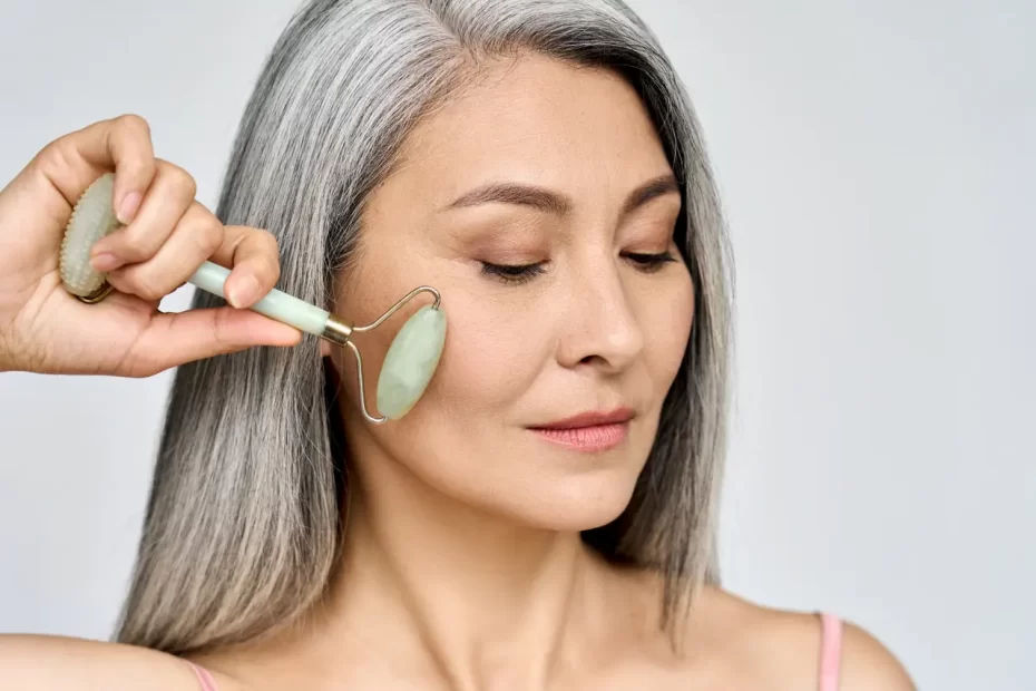
Repetitive facial muscle activity causes its fibers to stretch over time, creating wrinkles on the surface of skin over time. Therefore, many of today’s aesthetic treatments focus on diminishing these lines by suppressing their formation at epidermal level.
Botox injections may only address part of the issue. If the depressor anguli oris muscle is overactive or underactive, its hyperactivity or weakness will cause corners of mouth to appear as though they’re permanently drooping – an asymmetry which neurotoxin injections like Botox can correct.
Treatment is safe for adults and is generally performed at the doctor’s office by an experienced healthcare provider. Repeated injections may also contribute to muscle degeneration faster, so it’s essential that you find someone qualified who can perform this procedure efficiently and safely.
Muscle Innervation and Its Impact on Facial Expression
Depressor anguli oris muscle is a pair of triangular muscles located lateral to the chin on both sides, belonging to buccolabial group of facial muscles with other similar levator labii superioris alaeque nasi, levator labii superioris alaeque nasi, zygomaticus major zygomaticus minor orbicularis oris mentalis and incisive inferior muscles. They attach directly to mental tubercle of mandible and then connect directly to modiolus which attaches in angle of mouth drawing it downwards expressing sadness or questioning expression.
This muscle receives motor innervation from the marginal mandibular branch of facial nerve CN VII. For its vascular supply, the inferior labial artery of the face, which arises from maxillary division of facial artery provides it. Certain fibers continue below their origin point at mental tubercle to form transversus menti muscle (also referred to as mental sling).
Hypoplasia or Agenesis of this muscle is relatively uncommon and typically present from birth (congenitally). People living with it will typically have an asymmetric smile due to trauma or birth injury; alternatively it could be brought about due to neurological conditions like cerebral palsy, multiple sclerosis or stroke.
After years of repeatedly using our muscles to express emotions, our depressor anguli oris muscles can become shortened and develop visible grooves known as wrinkles. But treatment options exist such as Botox neurotoxins to help decrease muscle movement and thus diminish these lines.
Botulinum toxin treatment on this muscle was revealed by a 2010 study as a way of temporarily lifting corners of mouth and creating temporary neck lift. With far fewer risks than surgical procedures and lasting up to several months before effects fade away. Furthermore, it’s an inpatient procedure without hospital stay, making this approach suitable for those seeking temporary changes rather than long-term transformation of facial features.
The Function of Depressor Anguli Oris Muscle in Facial Expression
The depressor anguli oris muscle is a facial muscle responsible for moving the corners of our mouth downward and creating the expression known as frowning. It’s paired so there’s one on both sides of our faces; also connected to other muscles such as zygomaticus major/minor, buccinator, and risorius; as well as mixing with platysma muscle and cervical fascia.
Depressor anguli oris muscle fibers converge and run superiorly towards the angle of the mouth, where they meld with other lip muscles inserted by other muscle groups known as modiolus muscles. At this junction point they meet and interlace called modiolus.
Alongside the depressor anguli oris, there are other facial muscles that contribute to facial expressions. One such muscle is the orbicularis oculi – the circular muscle surrounding the eyes which closes eyelids and squints when closed; antagonizing this action are depressor anguli oris muscles which pull downward when someone smiles.
As you might guess, these muscles are connected by a network of tendons and fascia. Furthermore, they receive innervation from the same nerve CN VII.
These muscles control jaw opening and closing; the palatal muscle elevates corners of mouth when speaking; these do not form part of depressor anguli oris but still allow us to smile or frown appropriately.
Depressor anguli oris is a relatively small muscle, yet plays a large role in shaping facial features. When hyperactive, its contraction can result in excessive frowning expressions. Some individuals choose to inject it with botulinum toxin injections that block nerve signals causing it to contract, in order to improve results.
Understanding the anatomy of the depressor anguli oris can assist clinicians in diagnosing and treating conditions like CHDAOM more accurately. Kenhub’s detailed anatomy charts can offer more information about facial muscles.
Managing Asymmetry: Insights into the Depressor Anguli Oris Muscle
The depressor anguli oris muscle is a facial muscle located deep and laterally to the levator labii superioris muscle, bounding the infraorbital tissue space (canine space). Both muscles share innervation and blood supply from each other as well as from zygomatico-buccal muscle zygomatico-buccal muscle which runs along its bony attachments; furthermore they both share innervation from facial nerve (CN VII) via its branches zygomatic and buccal branches of facial nerve (CN VII), while maxillary Artery supplies blood to these two muscles as well.
The DAO muscle plays an essential role in facial expressions such as frowning and crying; its action plays a pivotal role. But as people age, this muscle becomes more likely to over-activate at rest causing its corners to drop down more often creating an indented or “gummy smile.” To combat this situation, many seek treatment such as Botox but keep in mind it requires periodic repeat injections for maximum effectiveness.
Congenital unilateral hypoplasia of the depressor anguli oris muscle in newborns often leads to an asymmetric crying facies; an uncommon clinical condition observed rarely. Pediatricians and otolaryngologists should seek early diagnoses by employing both clinical suspicion and an exhaustive search for anomalies elsewhere to ensure effective management of this rare disorder.
As well as its anatomical characteristics, the DAO muscle can cause various functional issues in the face due to its action. It has been linked with canine dental infections and TMJ dysfunction and reported as being at risk for accidental injection of columellar or alar branches of nasal arteries causing perioral hematomas. Furthermore, its action may be responsible for creating prominent “tongue grooves” or even pterygopalatocystic dystonia in some individuals.
After recovering from facial paralysis, one common sign is co-contraction between zygomatic and DAO muscles which obstructs smile. If this is suspected as being the case for you, a test using lidocaine in your DAO muscle could be conducted to assess its condition; to do this effectively you should seek assistance from an experienced otolaryngologist for this test.


