The External Rotators of Shoulder Exercise focuses on strengthening muscles like the Infraspinatus and Teres Minor, crucial for external shoulder rotation. By using resistance bands or light dumbbells, this exercise enhances shoulder stability, mobility, and overall joint health. Integrating it into a fitness routine contributes to improved strength and helps prevent shoulder injuries.
Your strength program should include exercises designed to strengthen and protect the shoulders. Two specific exercises targeting their external rotators – infraspinatus and teres minor) can increase performance while decreasing injury risks.
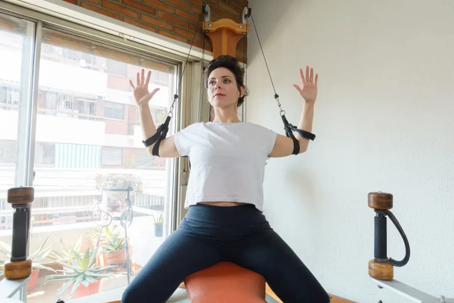
The TRA and ALT groups each participated in home-based progressive elastic resistance training of their rotator cuff muscles for eight weeks at home using progressive elastic resistance training, measuring maximal isometric strength of their left and right shoulder external rotators with the elbow flexed at 90 degrees for pretesting.
Infraspinatus
The infraspinatus muscle is one of the four muscles that comprise the Rotator Cuff in the shoulder. It is a thick triangular muscle that serves as an external rotator of the shoulder joint, helping produce shoulder extension. Originating on the medial (closer to spine) border of scapula it extends posteriorly towards glenohumeral joint before inserting on greater tubercle of head of humerus; innervated by suprascapular nerve and receiving arterial blood supply from circumflex scapular artery.
Infraspinatus is an integral part of the rotator cuff, playing an essential role in maintaining shoulder joint integrity and decreasing injury risk. Strengthening this muscle is critical to both shoulder health and reducing injury risk over time; otherwise, its tendon may weaken over time due to heavy loading causing more potential damage than strengthening exercises can address. For optimal shoulder health and injury prevention, exercises that target infraspinatus as well as other rotator cuff muscles should form part of any exercise program.
For infraspinatus strength training, prone horizontal abduction with external rotation is an excellent exercise option. This movement is often utilized in physical therapy sessions in order to increase shoulder mobility and prevent injury. Furthermore, this workout increases rotator cuff endurance; however it’s essential that the appropriate weight be used. Otherwise, overdoing it could lead to shoulder damage.
Standing Resisted External Rotation is another effective exercise for strengthening the infraspinatus muscle. It’s especially useful for athletes engaging in overhead activities like throwing and tennis; simply secure a band around a pole to allow your arm to rest at 90-degree abduction position – this activates both supraspinatus and deltoid muscles more heavily while stimulating your infraspinatus moderately.
A rolled towel can be placed between the elbow and patient’s side to reduce compensatory movements during standing resisted external rotation exercises, thus increasing infraspinatus activity while decreasing activation of other rotator cuff muscles. This modification creates greater activity for infraspinatus while attenuating other activation of other rotator cuff muscles.
Teres minor
The teres minor is one of four muscles forming the rotator cuff and is one of two prime movers for shoulder external rotation (along with infraspinatus). Together with its fellow members of rotator cuff it helps stabilize humeral head in shoulder socket. Even though it’s small in size it plays a vital role in shoulder movement and provides access to wide ranges of motion.
An exercise routine to stimulate it could include dumbbell side lying external rotation, face pulls, lateral raises and YTWLs. Prior to engaging in these exercises it is wise to warm up and stretch in order to loosen up muscles that become compacted over time – which could potentially lead to injuries. Also it would be prudent to minimize time spent typing, playing video games or using smartphones that put pressure on internal rotation of shoulders (this includes typing, video gaming or smartphone usage).
Teres minor is also essential in shoulder adduction – which refers to moving the arm towards its midline of body to reduce angles between arm and torso – with infraspinatus and teres minor also aiding this movement.
The teres minor muscle can be easily palpated at the infraspinous fossa of the scapula, while also being felt as an undulating mass when abducting your shoulder.
Contrary to its counterpart, the teres minor does not initiate shoulder abduction; it acts throughout the range, though less at 90 degrees than 30 degrees of abduction. Furthermore, its lower insertion point makes it an essential muscle in stabilizing shoulder stability.
People suffering from upper crossed syndrome (UCS) typically exhibit inhibited and lengthened teres minor muscles, which leads to their shoulder being rotated internally, leaving this muscle inactive and chronically stretched out. Implementing shoulder exercises targeting this muscle with proper technique will help alleviate this condition while simultaneously strengthening rotator cuff overall and improving performance of shoulder.
Posterior deltoid
The posterior deltoid muscle is the largest and triangular-shaped muscle overlying the shoulder joint, giving the shoulder its distinctively round form and acting primarily as an abductor, though also contributing to flexion and extension. This muscle is vitally important for anyone performing repeated overhead movement or carrying heavy loads; injuries to it can cause pain and weakness in the shoulder, making its development all the more important for professions that involve continuous overhead activity such as swimmers or pitchers.
The deltoid muscles are responsible for the adduction and extension of arms away from the body; its rear portion, however, extends them in an outward fashion. They play an essential role in many exercises including shoulder presses and dumbbell flyes; studies have also indicated that its lateral head contributes more towards shoulder flexion than extension as well as providing assistance to other transverse extensor muscles such as infraspinatus and teres minor.
Electromyography was employed in a study designed to assess the contributions of anterior, medial, and posterior deltoid muscles to shoulder movement by researchers using electromyography (EMG). Subjects performed five distinct shoulder exercises randomly with warmup on an upper-body arm ergometer before EMG measurements were collected at maximum voluntary contraction for each exercise – making an MVC determination possible for every EMG reading taken during this session.
Deltoid injuries occur when muscles become overstretched or overused. Overuse is especially risky during overhead activities like swimming and throwing a ball and can result in pain and swelling within the muscle itself. Treatment options for such an injury include rest, ice packs and heat applications as well as exercises prescribed by physiotherapists in order to strengthen and protect it from further damage.
Rear deltoid machines provide an effective and safe method of targeting posterior deltoid muscles, similar to rowing machines but specifically tailored to target these shoulder muscles. To use one, sit on its seat facing its pad while adjusting its height so the handles in front are at shoulder level; move arms backward as though swimming or pitching a ball and hold for several seconds before returning back into starting position and repeat until reaching your desired number of repetitions.
Extensor carpi radialis brevis
Extensor carpi radialis brevis is one of the seven muscles in the posterior compartment of the forearm, located beneath its elbow joint. Originating at the lateral epicondyle of the humerus along with two other muscles of this compartment and inserting at medial surface of ulna, innervated by radial nerve, and shorter and thicker than its counterpart Extensor carpi radialis longus it shares tendon attachment to abductor pollicis brevis and pronator quadratus attached directly onto extensor retinaculum on dorsal side.
ECRB users typically suffer from an overuse injury known as Lateral Epicondylitis (LET). This overuse injury occurs when the forearm extensor muscles become painful and tender over the lateral epicondyle of humerus due to repetitive use. Athletes and manual laborers who employ their hands regularly in repetitive activities, such as constant elbow flexion/extension, forearm supination, heavy lifting or wrist bending/twisting are susceptible.
Exercise to strengthen and relieve ECRB pain. Eccentric exercises on the ECRB may increase its shear elastic modulus and relieve tension on tendons; such exercises should be conducted with elbow in extension, forearm pronated, and wrist flexed toward ulnar deviation.
Patients suffering from LET should seek medical advice as soon as symptoms appear. Conservative treatments, including rest, activity modification, icing, NSAIDs and bracing may be tried first; if these measures prove ineffective then an MD may suggest more aggressive steroid injections or other noninvasive solutions.
The radial nerve is the primary blood supply to the ECRB muscle. It provides nutrients and oxygen during activity or stretching via its supply via the Ulnar Collateral and Radial Recurrent Arteries in the forearm, while also passing through the posterior cord of the Brachial Plexus where it’s protected from being pinched against structures by abductor Pollicis, Extensor Digitorum and Extensor Reticulum muscles protecting it during movement.


