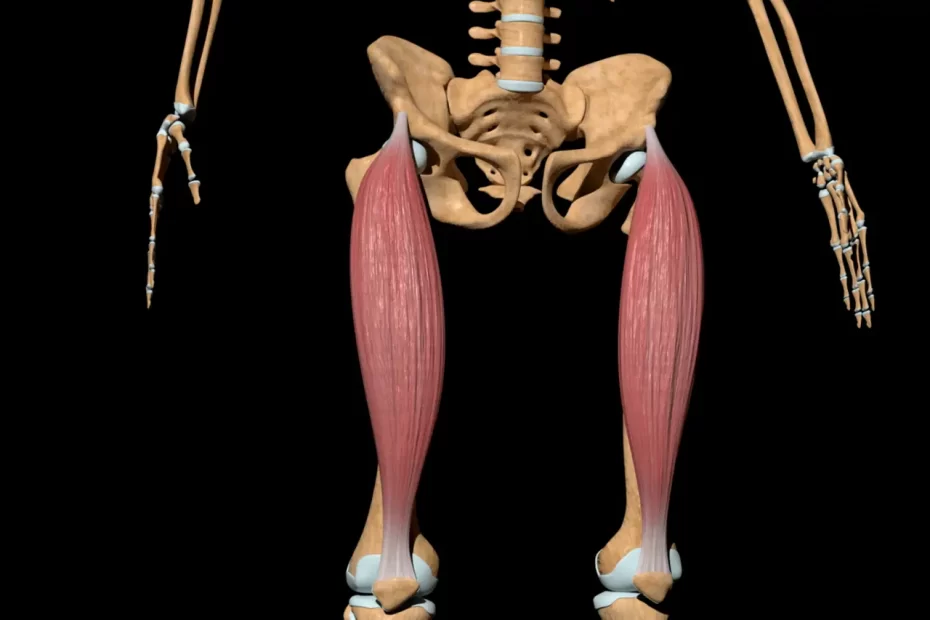
The rectus femoris muscle is one of the four quadriceps muscles, and serves to flex both hips and extend both knees. Its action involves flexing at the hip joint while extending both knees; its endpoint forms a broad and thick aponeurosis covering two-thirds of its posterior surface and eventually narrowing into a flattened tendon that inserts itself at the base of patellar bone.

Posterior femoral artery
Femoral Artery (FA): an essential blood vessel carrying oxygen-rich blood from your groin to leg. Due to its wide diameter, surgeons can use a catheter (thin flexible tube) easily access other blood vessels within your body. Femoral Artery serves as an important landmark for endovascular procedures like Percutaneous Transluminal Coronary Angioplasty (PTCA) or Stent Placement. The Femoral artery carries blood directly to your lower leg and comprises three layers. The tunica intima regulates blood pressure and prevents blood clots, while elastic fibers help your arteries expand and contract as they carry blood through them. Meanwhile, tough and durable tissues form an outermost protective barrier over your femoral artery to shield it from injury or damage.
The posterior femoral artery is the branch of the femoral artery that runs along the posterior wall of the rectus femoris muscle in the femoral triangle and often remains concealed by muscle and fascia. It branches off five times into this triangle as well as one branch going directly into adductor canal. Closely related to both femoral vein and pectineus muscles in its upper parts as well as passing in front of Sartorius muscle before passing under connective tissue covering known as adductor canal.
The posterior femoral artery contains multiple nerves in addition to its main vessel; these nerves control movement of the quadriceps femoris muscle, composed of four muscles between hip and knee joints that cover gait from flexion of hip flexure to extension during gait. Together with iliopsoas muscles, quadriceps femoris acts together with its partner the quadriceps femoris (translated: four-headed muscle).
The Femoral Artery is an extension of the External Iliac Artery that runs medial to the Inguinal Ligament at the Inguinal Crease. It then flexes to follow the Femur inferiorly and medially before ascending posteriorly through Hunter’s Canal as Popliteal Acheron for posterior knee, then calves, foot as the Popliteal Artery for lower leg. Hunter’s canal is commonly referred to by its acronym.
Femoral nerve
The femoral nerve provides blood and nutrients to the rectus femoris muscle, as well as controlling its counterpart: quadriceps femoris, which extends and flexes knee joints. Any injuries to quadriceps femoris can come about from several sources but usually involve its tendon, connecting inner to outer muscles of knee. Most quadriceps injuries are minor and can usually be treated through rest, ice packs and rehabilitation exercises.
The anatomy of the rectus femoris is complex, and was only fully understood in 1995. This muscle has two tendinous sources – one from AIIS (anterior inferior iliac spine) and the other from SAR and PLC (6a). They converge 2 cm distal from their points of origins to form one tendon about 2 cm distal; although close by, each retains its individual identity with only 10-20% fibers merging distally.
MRI imaging has proven itself useful in the identification of rectus femoris musculotendinous injuries. Such injuries include inflammation, avulsion and/or fracture of an aponeurosis or muscle belly; injuries often arise during intense activities like running and jumping and may range from partial tears or ruptures all the way through to complete ruptures of this important muscle group.
Symptoms of rectus femoris strain include pain in the front of the knee and a clicking or popping sensation when trying to bend your leg at the hip. A rectus femoris avulsion fracture may cause swelling, bruising and tenderness in quadriceps area of thigh; such injuries can be seen using an MRI scan or T2-weighted image.
Femoral nerve blocks involve inserting a needle with electrical stimulation devices into your patella, which will make it twitch and indicate to your provider they have reached the femoral nerve. Although painful, recovery from such procedures usually takes between four months to one year.
Medial femoral artery
The medial femoral circumflex artery (MFCA) is an essential source of arterial blood supply to the quadriceps femoris muscle. Running from between the iliopsoas muscle and pectineus, this vessel provides most of the arterial blood supply to the femoral head. Variations in its origin or course can sometimes result in serious medical complications, including arteriovenous fistula formation [1].
The quadriceps femoris is a powerful extensor of the knee joint in the anterior compartment of the thigh. Composed of four muscles – rectus femoris, vastus lateralis and vastus intermedius – this muscle forms the quadriceps tendon at the base of the patella. Surrounded by fascial expansion called the “femoral sheath”, which contains nerve, artery and vein connections for optimal performance, these four heads work as one unit in harmony!
This study investigated the anatomy of medial femoral circumflex arteries (MFCAs) in 342 cadavers (158 female and 184 male), with most beginning at the common femoral artery and running between pectineus and psoas major muscles before branching off to form terminal branches. An unusual variant originating from the femoral vein has also been noted.
We examined the relationship between the MCFA and the lateral femoral circumflex artery, which provides arterial supply to the knee joint. Most often, this arterial vessel (LCFA) runs parallel with MFCA and crosses just proximal to inguinal ligament. However, in rare instances it can pass proximal of MCFA to run along inferior border of Obturator externus towards intertrochanteric crest.
MCFA is also an important source for the insertion of the rectus femoris muscle, serving both blood and nerve supplies that provide functional reconstruction of thigh. Innervated by the femoral nerve, this method offers functional reconstruction; however it comes at the cost of loss of muscular strength as well as needing an extensive donor site that accommodates this musculature.
Medial collateral ligament
The Medial Collateral Ligan (MCL) is a thin band of connective tissue extending from the front of the femur to the medial aspect of the tibia and supporting the knee joint. As one of four major ligaments supporting it, it is commonly injured during sports like skiing and football; most frequently when skiing and football players take an aerial fall. Furthermore, its role as an essential tensor of quadriceps femoris tensor makes an MCL injury often resulted in patella dislocation after injuries to other major ligaments such as ACL injuries occur simultaneously with injuries to another major joint or another major joint supporting its support of course!
The superficial medial collateral ligament (sMCL) consists of two layers, the superficial layer and deep layer. It is especially vulnerable to injury during situations in which there are combined flexion and extension forces acting against it, such as during valgus loading. Therefore, understanding its stress distribution in terms of strain measurements at various flexion angles was undertaken for research purposes. These results demonstrated an elevated strain near its insertion point during valgus loading which suggests increased chances of rupture for this ligament.
Another finding from this study revealed a higher strain at the proximal part of MCL than its distal end, possibly as a result of it generating most of its strain during knee flexion phase, in contrast to SCL which only produces part of its force during this stage.
Studies into the MCL have been extensive, and numerous techniques exist for gauging its strength. Most commonly employed is magnetic resonance imaging (MRI), while CT scanning can show ligament thickness which serves as an indicator of its strength.
The rectus femoris muscle, part of the quadriceps group of muscles that support the knee, is a biarticulate muscle as it spans both joints in its path. Primarily used to extend knee joints further back than they’d otherwise go, this biarticulate muscle also assists iliopsoas muscles with hip flexion when needed and receives blood from its own descending branch within the lateral circumflex femoral artery for nourishment.


