Strengthening primary internal rotators of the shoulder with physical therapy exercises may help decrease impingement and enhance glenohumeral joint movement.
Flexion–Defined as the act of moving the upper arm forward along its sagittal plane. The main flexors for shoulder flexion include deltoid and coracobrachialis muscles.
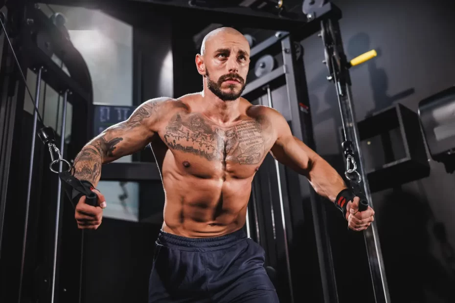
Rotator Cuff–The rotator cuff is composed of muscles that provide stability to the “ball” in its socket in the shoulder blade, controlling both shoulder rotation and lifting movements. It provides for efficient lifting motion.
Supraspinatus
The supraspinatus muscle is one of the four tendons and muscles that comprise your shoulder’s rotator cuff, each helping with different aspects of its movement while working collectively to hold your upper arm steady in its socket. The supraspinatus muscle is responsible for abduction movement away from your centerline (abduction). It provides 15 degrees of abduction before being gradually replaced by deltoid muscles. Subspinatus injuries often result from overuse or trauma to the shoulder, including falling on it, lifting heavy objects improperly, dislocating your shoulder joint, dislocating its joints and dislocating itself. A supraspinatus tear may either be acute or degenerative in nature – usually the latter, caused by repetitive activities that involve lifting arms above one’s head like painting and carpentry, may develop over time.
Importantly, it should be remembered that the supraspinatus muscle has both an anterior and posterior belly. Typically, the former tends to be longer and thicker than its counterpart; studies have also demonstrated that its activation during abduction contributes more directly to force coupling with deltoid.
Injury to the supraspinatus muscle can result in pain on both sides of the shoulder. This pain increases with abduction and elevation of arm movements and makes daily activities difficult; sometimes there may also be tenderness in upper trapezius muscle.
Professionals (PT, ATC, DC and OT) can assist in alleviating shoulder pain by palpating and assessing the supraspinatus muscle tone. Manual mobilization techniques such as anterior to posterior and superior to inferior movement may help decrease tension in this muscle group.
If you suffer a supraspinatus injury, consulting with an orthopaedic surgeon as soon as possible to evaluate treatment options is vital to recovery. Your physician can evaluate your condition and suggest either conservative rehabilitation or surgery as the optimal course. At Thomas & Bigler Knee & Shoulder Institute in Las Vegas and throughout Nevada, board-certified orthopedic surgeons treat supraspinatus injuries.
Infraspinatus
The infraspinatus muscle is one of the rotator cuff muscles and, like its counterpart, helps flex the shoulder. Additionally, it contributes to lateral rotation of the arm and plays an active role during activities like climbing or throwing a ball. Innervated by the suprascapular nerve at roots C5 and C6, its arterial blood supply comes from superior and circumflex scapular arteries.
The Infraspinatus muscle can be found on the dorsal surface of the scapula and lies deep to both trapezius and deltoid muscles, often being mistaken for its counterpart the Teres Minor Muscle since their fibers run parallel, only separated by spine of scapula spines. Its tendon inserts into inferior facet of humeral tubercle, providing second strongest anatomic force against lateral rotation of shoulder (after subscapularis).
At higher abduction angles, Infraspinatus does not generate external rotator forces as effectively due to its decreasing moment arm as abduction angle increases; however, it provides great external rotation at lower abduction angles and can help stabilize shoulder stability for activities such as throwing, washing hair or reaching for items over head.
Muscle function is integral for full range shoulder movement and should be assessed in all patients suffering from shoulder pain. This should involve testing in multiple positions in order to ascertain which muscles are active for each motion. In order to do a complete inspection of shoulders and detect signs such as swelling, asymmetry, winging of the scapula (winging) or muscle atrophy it may also be helpful to disrobe for examination of all parts of this joint system.
Apley scratch test is an effective way of measuring shoulder external rotation. Patients should place their arms by their sides without touching their trunk with elbows flexed at 90 degrees; after which an examiner will stretch out forward and upward against resistance in order to assess shoulder external rotation. More flexion occurs in this maneuver indicates greater internal rotator strength in the shoulder.
Teres Minor
The Teres Minor Muscle is part of the Rotator Cuff and one of the intrinsic shoulder muscles (scapulohumeral internal rotators). Originating at the lateral border and posterior surface of the Scapula and inserting on to greater tubercle of Humerus. Innervated by Axillary Nerve; provides part supply to Posterior Circumflex Humeral Artery.
Reducing or weakening the teres minor muscle can result in shoulder pain and reduced internal rotation, instability in shoulder position and upper crossed syndrome. To restore proper function to this muscle it’s key to improve shoulder stability which can be accomplished using scapular stability exercises.
Before any workout involving overhead movement of the arm, it is crucial to release and stretch out the teres minor muscles by performing several sets of prone arm-supported internal rotations, similar to what can be seen above.
Alternately, you can do the same exercise by placing your hands in front of your body while moving your shoulders into a retracted position against resistance. Complete 2-3 sets with 10-15 repetitions each.
Work the teres minor by engaging in shoulder movements that require some degree of external rotation, such as clean, snatch, pull Olympic lift variations or dumbbell/cable bicep curls with external rotation incorporated. However, be wary of overtraining it since its contraction leads to only minimal scapular plane adduction during maximal contraction.
The teres minor muscle is located beneath both infraspinatus and subscapularis and provides shoulder stabilization during external rotation, especially during external rotation of external rotation. Its fibers run superolaterally toward the greater tubercle of humerus in a radiate pattern to form its radiate muscle shape; its fibers converge at this greater tubercle to form its radiate shape and play an essential role for shoulder stabilization and rotator cuff function, particularly external rotation; similarly like supraspinatus its activation can be inhibited by shoulder internal rotator stiffness; so addressing internal rotator stiffness may help increase teres minor adduction at the shoulder joint.
Subscapularis
Located in the subscapular fossa, the Subscapularis muscle is one of four muscles that form the rotator cuff with the Supraspinatus, Infraspinatus and Teres minor. The rotator cuff muscles help to steer and stabilize the head of the humerus within the glenohumeral joint. The primary function of the Subscapularis is to perform internal rotation of the shoulder.
The muscle fibers originate in the subscapular fossa of the scapula and attach to the medial lip of the bicipital groove on the humerus. A broad tendon accompanies the middle third of the muscle, which inserts on the lesser tuberosity of the humerus. The footprint of the subscapularis tendon on the lesser tuberosity has been described as consisting of four to six thick collagen bundles (Fig. 30-10).
Like the other rotator cuff muscles, the Subscapularis can be injured. A direct injury such as falling onto the outstretched arm or a sudden traumatic contraction can cause tearing of the subscapularis tendon. Indirect injuries may occur from repetitive motions in jobs or sports that require repetitive shoulder actions with forceful internal rotation, such as baseball pitching and tennis. This may lead to overload of the muscle-tendon complex, resulting in a strain response with subsequent fibrosis and fatty tissue deposition within the tendon, causing tightness and weakness.
A partial or full-thickness tear of the Subscapularis tendon may cause pain and weakness in the shoulder. On physical examination, the patient will have pain localized over the lateral aspect of the shoulder and a loss of external rotation power (Fig. 31-10). A positive Neer test is usually present.
Isolated subscapularis tears are uncommon, but they can be difficult to diagnose and have a slow onset of symptoms. They can be associated with pain localized over the lateral shoulder and with referred pain down the biceps brachii tendon to the front of the elbow. They can also be associated with difficulty with overhead activities and weakening with external rotation of the shoulder. They can be diagnosed by performing a lift-off test against resistance, first described by Gerber and Krushell. The patient is asked to extend, adduct and internally rotate the shoulder and to lift the hand off against resistance.


