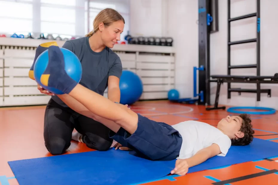
Abductor muscles work to move legs away from pelvis through an action known as adduction, making these an integral part of any workout program. Strengthen these important muscles with targeted abductor exercises or seek advice from an exercise physiologist or physiotherapist for guidance.
Groin pain from hip abductor muscle tears is often hard to diagnose and treat effectively, but Dr Okoroha may recommend physical therapy as a means of relief.
The Rectus Abdominis
The Rectus Abdominis muscle is a long and thin structure located along the anterior abdominal wall that gives people with very low body fat a “six pack” appearance. Along with internal oblique and transversus abdominis muscles, it forms the core muscle group. When activated it flexes trunk anteriorly before working together with other abdominal muscles to compress abdomen increasing intraabdominal pressure (critical during forced breathing, labor, defecation and micturition). Furthermore it controls pelvic tilt (antilordosis), controls pelvis tilt (antilordosis) while helping stabilize spine movements by stabilization of spine stabilization during movement.
The muscular structure of rectus abdominis can be described as being composed of horizontal fibrous bands called tendinous intersections, connected by tendinous ligaments. One tendinous intersection usually exists near the umbilicus; another at the inferior tip of xiphoid process and third one located midway between. Innervating this muscle are three nerve branches from T7-T11 intercostal nerves which penetrate its fascial covering called the thoraco-abdominal nerves which innervate this muscle.
A wide range of other structures exist within the rectus sheath, such as the inferior epigastric artery and vein, lymphatic vessels, and the aponeuroses for external oblique, internal oblique and transversus abdominis muscles. Furthermore, this sheath receives contributions from six lower intercostal nerves via segmental contributions.
The Rectus Abdominis is the largest of the flat abdominal muscles, located on either side of the spine. It arises from both front lateral pubic tubercle and anterior portion of iliac crest and inserts into linea alba at lower edge of sternum and costal cartilages of ribs 5-7; some fibers of Rectus Abdominis may extend lateral to fourth and fifth ribs but this slipage usually isn’t visible.
Like its abdominal counterparts, the rectus abdominis must be strengthened through exercises which flex and stabilize the spine, such as crunches, planks and movements like farmer’s walks, dead lifts, prone dumbbell rows and pallof presses. For optimal development of this muscle group, training 2-3 days each week with 8-10 reps should suffice.
The Adductors
Adductor muscles – composed of pectineus, adductor longus, gracilis and adductor magnus muscles – are inner-thigh muscles responsible for drawing the legs towards the center of your body while walking or running. They help maintain hip position while creating adduction torque (bringing legs closer together); additionally contributing to knee flexion making these adductors hip flexors as well.
Adductor muscle groups are attached to the pubic bone just like abdominal muscles (your six-pack), meaning when one muscle is injured it usually affects both sets. Thus a problem with your abdominals could also lead to inner thigh discomfort – making training both groups at the same time especially important.
Adductor insertion points are situated very close to those of rectus abdominis tendons, making it easy for these two muscles to work in concert during athletic activities, often leading to injuries on both sides. Tendons of both muscles often suffer injuries at once.
From its front view, the adductor magnus appears more like a single-joint hamstring than a quadriceps muscle and can help protect your pelvis and upper body from leaning forward just as efficiently as its counterparts hamstrings. From its rear view however, however, its appearance drastically changes; from here you’ll notice it has a wide base that extends along the length of the femur before fastening securely onto medial epicondyle (a bump on inside your knee).
Adductors can be difficult to target as beginners, so the side-lying leg lift exercise is an effective starting point. To perform it, lie on your side with one leg bent and foot flat on the floor in front of you; lift your bottom leg off of it using adductor contraction before slowly lowering it back down onto its previous position; repeat for 10-12 repetitions or more to build strength in adductor muscles.
The Transversus Abdominis
The transversus abdominis muscle is one of the deep abdominal muscles, forming a horizontal band of tissue along your abdomen. Its free lower border lies at the deep inguinal ligament, where it communicates with both internal and external oblique muscles. Connected to both spine and pelvis by nerves such as intercostal nerves, the iliohypogastric nerve and ilioinguinal nerve.
Transversus abdominis serves to stabilize and support your torso by maintaining regular abdominal wall tension, thus preventing unnatural back movements that could otherwise occur from movement of lumbar spine and pelvis.
It also plays an integral part in protecting internal organs by holding them in place and compressing them, as well as providing dynamic stabilization – which means stabilizing core movement during movements and posture changes.
Other functions include withholding bowel movements and aiding labor contractions and pushing during childbirth. In addition, this muscle tightens during a Valsalva maneuver in which your thorax tightens when you hold your breath causing your sphincter unknowingly tightening to help support breathing (11-16).
As it lies deep, this muscle is harder to identify and engage than other core muscles, but can still be strengthened through core strengthening exercises like string vacuum. To perform it properly, lie face down on a floor mat with legs together and arms straight above you before pulling them up to 12-18 inches (30-46 cm). This exercise targets transversus abdominis muscle by pulling it in and contracting it for an extended period. Other exercises which strengthen it include supine leg lifts and hollow body holds.
Strengthening abdominal muscles can lower your risk of hernia by increasing their capacity to contract and provide support to abdominal organs. Furthermore, properly performing core strength exercises may decrease injury risks when moving or engaging in typical postures; an allied health professional can teach proper “core strengthening” techniques designed to strengthen both your spine and pelvic stability.
The Rectus Femoris
The rectus femoris muscle runs down the front of each leg as part of the quadriceps group and extends and flexes both knees and hips. A rupture of its tendon can occur through repetitive knee extension movements like sprinting or kicking, as well as during explosive jumps – often leading to pain in the groin and difficulty raising legs or knees; risk factors for this injury could include poor hip or knee mobility, inadequate warm-up sessions and fatigue of muscle fibers.
MR imaging features of the rectus femoris muscle include its fusiform appearance with two tendon sheaths that encase its belly. It has a wide posterior aponeurosis that joins with vastus medialis and vastus intermedius tendon to form quadriceps tendon; from there its tendon connects directly to patella via quadriceps ligament before inserting onto it at base of patella via quadriceps ligament and inserts onto it directly. It also features small aponeurosiss that inserts directly into both ilium and pubis muscles;
Due to its unique muscle within a muscle architecture, the rectus femoris is vulnerable to tears and strains, which can be difficult to diagnose. A common injury pattern involves when inner bipennate muscle becomes separated from overlying outer unipennate muscle at proximal musculotendinous junction, making this injury hard to distinguish from an avulsion fracture of tendon sheath.
On fluid-sensitive sagittal MRI images, a tear at the proximal musculotendinous juncture can be seen, with its outer layer of muscle and deep middle portion that contains an intramuscular cyst. Another injury pattern seen is partial dissociation of inner bipennate muscle – visible with a distinct gap between its fibers and surrounding fat tissue on fluid-sensitive images.
Rectus femoris tendon rupture is often seen in sports that involve repetitive knee extension motions such as running, jumping and kicking; it is also often observed among athletes participating in explosive movements like sprinting, martial arts and football. Diagnosis can be made by taking a history of injury as well as conducting physical examination and ordering diagnostic tests such as ultrasound imaging, magnetic resonance imaging or x-rays; treatment options include rest, ice compresses as well as rehabilitation or physical therapy for rehabilitation of physical therapy treatment options for recovery of physical therapy treatment options for rehabilitation/physical therapy purposes.


