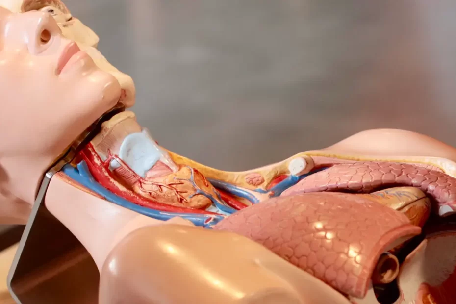
The posterior belly of digastric is one of two bellies comprising this muscle. Located above the hyoid bone, its supply comes via the digastric branch of facial nerve.
There have been a variety of variations affecting the anterior belly of digastric. Some are quite fascinating; we will discuss a few in this article.
Origin
The digastric muscle is a small pair of paired muscles located in the anterior compartment of the neck. It belongs to a group known as suprahyoid muscles that also includes mylohyoid and geniohyoid, with two muscular bellies (antero and posterior). Each belly has different embryological origins and thus innervations – the posterior belly of digastric receives nerve supply from facial nerve CN VII while mylohyoid receives innervation via inferior alveolar nerve.
The posterior belly of the digastric begins at an indentation on the lower surface of the skull medial to the mastoid process of the temporal bone known as the mastoid notch or digastric groove and extends ventrally and inferiorly into an intermediate tendon of stylohyoid muscle, while its anterior counterpart originates on its superior surface from a similar indentation; both bellies of digastric are connected via an intermediate tendon connected to body and greater horns of hyoid bone.
When activated, the posterior belly of the digastric elevates or depresses the hyoid bone to initiate opening or closing of mouth. This muscle plays an essential part in this function and in normal function is assisted by both mylohyoid muscle movement and sternocleidomastoid muscle lateral movement.
This muscle serves as an anatomical landmark to help locate the courses of accessory nerves, internal jugular veins and carotid arteries that must be preserved during neck dissections. Furthermore, its attachment point for the phrenic nerve can also be easily identified using this landmark.
At times, an abnormal anterior belly may be found in muscles. This typically consists of an accessory muscle originating from the digastric fossa that inserts into its intermediate tendon; Quadros et al reported one right accessory anterior muscle from this source insertion directly into its second anterior belly; they also observed one from mylohyoid raphe originating in that same antrum and inserting directly into its right intermediate tendon of digastric.
Insertion
The digastric muscle is a bilateral muscular structure in the neck. It consists of thick and strong fibers which stretch from the front of the oesophagus to the lower border of the hyoid bone. Served by facial nerve, this muscle plays a part in deglutition. As one of the largest of pharyngeal muscles with two bellies located anteriorly and posteriorly joined together via an intermediate tendon.
Neural crest cells migrate caudally during embryogenesis to form five pharyngeal arches which provide muscle-forming tissue and their derivatives, including digastric muscles whose anterior belly derives from arch one and is supplied by mylohyoid nerve, while its posterior belly comes from arch two and supplied by facial nerve.
The anterior belly of the digastric begins at the lateral surface of the mastoid notch of the temporal bone and travels toward the hyoid bone, passing close behind (posterior) the stylohyoid muscle before becoming attached to both its body and greater cornu by an intermediate tendon.
The posterior belly of the digastric muscle lies within the submental triangle and is directly associated with the hyoid bone. Furthermore, its reach extends to include the omohyoid bone at the base of throat where it connects via the peripalatine ligament to this small bone in its center. Furthermore, its proximity lies close to internal and external carotid arteries, deep cervical lymph nodes, jugular veins, vagus nerves, and hypoglossal nerves.
De-Ary-Pires et al. [9] reported an absence of posterior belly in one side while Ozgursoy and Kucuk [31] described bilateral posterior accessory bellies of the digastric muscle; Quadros et al.[38] reported on an accessory muscle bellies with varied origins and insertions that originates in the digastric fossa and inserts into second anterior belly of digastric muscle. Variants in anterior bellies are more prevalent; variants in posterior belly are less frequently reported than variants reported with regards its origin or attachment points to second anterior bellies of digastric muscle.
Nerve Supply
The digastric muscle is normally innervated by the trigeminal nerve via its mylohyoid branch and comprises two structural bellies; an anterior belly (AB) and posterior belly (PB), connected by an intermediate tendon that perforates stylohyoid muscle and secured with a fibrous loop that may or may not have a mucous sheath.
Variations in the anatomy of this muscle are not unusual. Variations usually involve its origin and insertion. There have been reports of other abnormalities as well, for instance the “real quadrigastric muscle”, with three accessory anterior bellies that originate at the lower border of the mandible and insert directly into its intermediate tendon without crossing it; another variation includes two accessory muscles from midline raphe insertion to its respective PB (posterior belly muscle).
The anterior and posterior bellies of the digastric muscle receive their arterial supply from the submental artery of the facial artery, running between mylohyoid and submandibular glands. Although anatomic variations can exist in how these arteries supply these muscles (Faltaous and Yetman’s report indicates this, for instance), sometimes running shallow instead of deeply into its vicinity – as evidenced in 50% of cadavers studied!
There have been reports of variations in the nerve supply to the digastric muscle. For instance, cases may exist where its anterior belly is supplied by both facial and mylohyoid nerves; furthermore it’s possible for one additional twig derived from nerve to splenius capitis to supply its posterior belly on the left side. These abnormalities should be taken into consideration when reading MRI/CT scans of the neck as well as when performing surgical procedures in this region as failing to recognize these changes can lead to misdiagnoses as well as surgical complications.
Complications
The digastric muscle is an integral component of swallowing and speech production, helping with mandible depression and elevation of hyoid bone to allow swallowing and speech production, posterior tongue pulling during chewing and biting, trigeminal nerve innervation from its anterior belly (cranial nerve V) as well as facial nerve innervation (cranial nerve VII), with mylohyoid nerve innervating its inner surface on its anterior belly belly.
The muscle has two bellies – anterior and posterior ones – connected by an intermediate tendon running through the stylohyoid muscle. Each belly receives input from different branches of facial nerve – mylohyoid branch and submental branch of facial artery respectively – while each belly receives blood from their respective source (mylohyoid branch versus submental branch of facial artery, respectively). The anterior belly appears thin and fleshy while its counterpart posterior one appears more flattened and compressed compared to its counterpart.
Numerous structural variations of the digastric muscle have been described in literature, most often regarding shape and location of muscle attachments. Furthermore, variations exist regarding insertion and innervation for each belly.
Cadavers demonstrate that in 70% of cases, the submental artery runs deep to the anterior belly of the digastric muscle while superficially in 30%; additionally, the anterior belly receives innervation from mylohyoid muscle innervation which makes it more vulnerable to changes in its anatomy.
Posterior bellies are more susceptible to injuries than their anterior counterparts, which may occur as the result of trauma or other factors. An injury may cause difficulty swallowing, hoarseness and neck or jaw pain as well as damage to an intermediate tendon which occurs from opening one’s mouth for extended periods or while chewing food.
Thankfully, the posterior bellies of the digastric muscle can be restored over time with relaxation exercises or injections with BTX-A injections to restore muscle function.


