Muscle contraction can either pull the pelvis forward, or rotate the hips laterally – hip lateral rotation being an example.
Recent research into the deep six hip external rotators produced high-quality muscle architecture data using cadaveric specimens and normalized measurements based on sarcomere length. This allowed each muscle to be identified independently from joint position or other factors impacting on muscle-joint kinematics.
Gluteus Maximus
Gluteus Maximus is the outermost and largest gluteal muscle, creating a thick, fleshy mass in your buttocks and commonly referred to as the “gluteal muscle.” As well as providing primary external rotation of your hip, Gluteus Maximus acts as an important hip stabiliser, controlling pelvis position during movement and helping control hip rotation during movements.
Studies have demonstrated that the lateral gluteal muscles (gluteus medius and minimus) can perform simultaneous hip abduction and internal rotation torque; this ability marks human bipedalism as it allows humans to simultaneously abduct and rotate the hip.
Functionally speaking, hip external rotators are at their most efficient when one leg bears weight over another limb during walking and running – an action which involves both planting and cutting forward motion with one action – something best executed by gluteus maximus, an organ specifically tailored to perform this action.
Viewed from above, hip muscles lie posterior-lateral to their joint’s axis of rotation (see FIGURE 1). Primary external rotators include gluteus maximus, obturator internus and externus, sartorius pectineus and long head of biceps femoris as primary external rotators; additional muscles that contribute include quadratus femoris and iliopsoas, but these have less of an impact due to small moment arm length and relative cross sectional area contributions than their primary counterparts do.
Gluteus Medius
The gluteus maximus tends to garner much of our focus when discussing weight-bearing hip movements such as squatting and running; however, its smaller triangular cousin — gluteus medius — should not be neglected when investigating hip lateral rotation. Gluteus medius is responsible for abduction, internal/external rotation of the hip as well as stabilizing pelvis during weight-bearing activities.
Locate your gluteus medius by placing your hand over one of your hip bones (iliac crest). As you abduct one leg at a time, slowly abducting will cause its muscle under your hand to contract and start contracting as part of its long tendon from behind your waist bone (ilium) which connects directly to the outer bony outcropping called greater trochanter through which this muscle connects via tendon attachments.
As with other short external rotator muscles, the gluteus medius is specifically tailored to produce an effective amount of hip lateral rotation torque when contracted – its force vector intersects nearly vertically with that of hip’s sagittal plane of rotation when this muscle contracts.
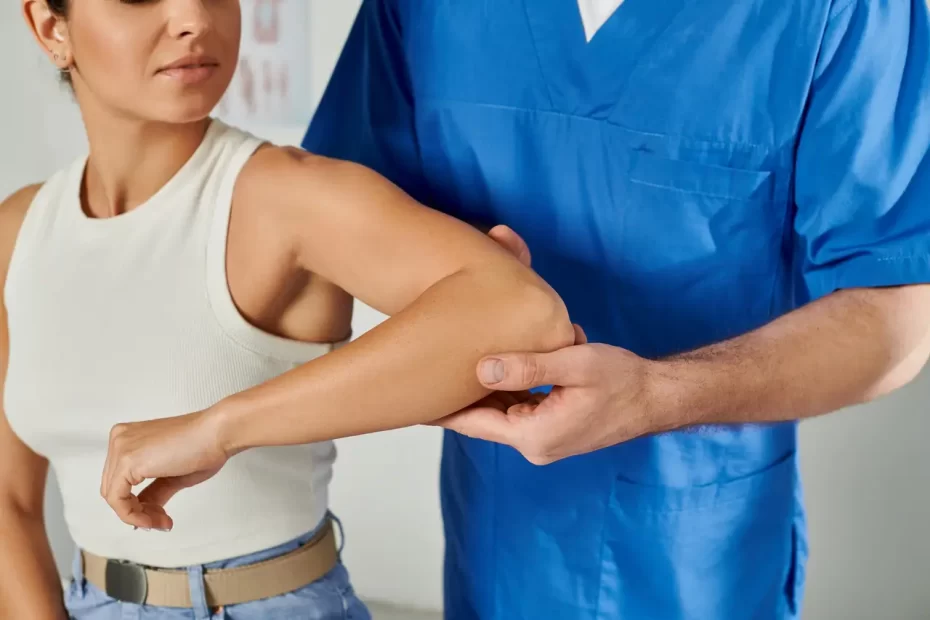
Physical therapists can screen for weakness in these hip external rotator muscles and prescribe strengthening exercises to avoid injuries to this area of the hip joint. When weak gluteus medius muscles and other short external rotator muscles cause instability or excessive hip rotation during weight-bearing activity, leading to instability or overrotation that could potentially result in injury. If necessary, physical therapy services could prescribe strengthening exercises designed to avoid such incidents from occurring in order to help avoid them from leading to potential instability and excessive hip rotation during weight bearing activity resulting in injury; physical therapy practitioners would be able to assess weaknesses within this muscle grouping as well as suggest strengthening exercises intended to strengthen them so as to help avoid them from becoming injury by screening them for possible weakness in these muscle groups while prescricing strengthening exercises in order to help avoid such incidents from occurring in order to ensure such injuries being avoided in order to help avoid such incidents from happening occurrence by screening them for signs and then prescricing strengthening exercises for these hip external rotator muscle weakness in order to prevent such injuries occurring from happening. Physical Therapists can screen these hip external rotator muscles as well as provide strengthening exercises in order to protect these injury occurring due to weakness within them and provide strengthening exercises in order to protect from happening by way of injury prevention exercises in order to prescribe strengthening exercises which help strengthen them before being injured as soon as possible and prescribe strengthening exercises as prevent any such injuries from happening in future situations.
Gluteus Minimus
Even though they may not be as widely recognized as other muscle groups such as gluteus maximus, medius, and piriformis muscles, deep six lateral hip rotators play an integral part in hip actions such as rotation. Together with sartorius pectineus obturator internus and externus they form part of what contributes to lateral rotation in hips.
The gluteus minimus starts at the posterior superior iliac spine and inserts into the iliotibial tract which connects with the greater trochanter lateral face. Unfortunately, this muscle can often shorten too quickly due to adaptive shortening or overactivity; stretching it is essential in maintaining its health and functioning effectively.
This muscle can provide both hip abduction and internal rotation torque, which is essential for running and other forms of movement. Furthermore, it plays an integral role in pelvic stability by stabilizing the opposite hip during gait.
Additionally, the gluteus minimus may invest in the anterior hip capsule and ischiofemoral ligament, explaining why its insertion is one of the primary sites for trochanteric bursitis as well as hip abduction pain.
Obturator Externus
The Obturator Externus (OE) is another hip muscle that assists in lateral rotation and adduction, receiving its blood supply via both the Obturator Artery and Medial Circumflex Femoral Artery.
Although important, this muscle is widely under-reported. Only one study was conducted, providing fiber length and physiological cross-sectional area (PCSA) measurements from one specimen used cadaveric specimens only; nevertheless it provides an excellent starting point for future analyses to predict muscle function.
This muscle originates at the external bony margin of the obturator foramen with a cylindrical tendon that passes under the neck of the femur to insert into the trochanteric fossa. Its primary action is hip external rotation; however, it also assists with hip adduction and pelvic tilting.
The obturator externus lies along the lower margin of the acetabulum and in neutral position contacts its superior portion; during abduction or external rotation this muscle touches more directly upon its inferior aspect, receiving innervation from the obturator nerve.
Sartorius
The Sartorius Muscle is unique because it spans both hip and knee joints. This action creates a cross-legged position commonly utilized when sitting or standing, which creates opportunities to flex, abduct and externally rotate the knee joint.
The muscle originates at an anterior superior iliac spine (ASIS) of the pelvis. From there, its fibers travel inferomedially until reaching their final destination at medial aspect of proximal portion of tibia. Femoral nerve innervates this muscle, with connections from spinal nerves at levels two, three, and four providing it with innervation.
Sartorius receives its blood supply from multiple arteries which penetrate it at irregular intervals; these include the superficial circumflex iliac, lateral femoral and deep femoral arteries. Due to its wide range of actions and span across both hip and knee joints, the sartorius muscle can easily sustain injuries that threaten its functionality and cause strains, tears or pes anserine bursitis. Common examples are hip flexor strains, extension tears or pes anserine bursitis. These injuries typically affect athletes participating in sports that involve frequent twisting movements around their planted leg, like basketball and football, as well as repetitive activities like long distance running and squatting. Common symptoms for this injury include burning sensation in front of hip and tenderness on inside knee.
Psoas
The Psoas Muscle (commonly known as Soul Muscle) is one of the most essential muscles in your body. Beginning at your lower back and moving alongside spine and ribcage to lesser pelvis brim where it joins up with Ilicus Muscle at base of Femur; they form ILIOPSOAs Muscle Group that plays an essential role in hip flexion as well as lower back movement during walking, running, bipedal loading.
Your psoas has one of the highest moment arms among all muscles in your body, meaning it can generate significant torque to move legs and hips. Unfortunately, however, its potential for movement is significantly weaker compared to other hip muscles; especially when in an anatomic position.
As such, it is crucial that your psoas muscle remains strong and flexible as possible if you engage in work that involves sitting for extended periods. Tight psoas muscles can lead to low back pain as well as other lumbar back issues like disc herniations or sciatica; regular practice of Clinical Somatics exercises may help release and strengthen it further.


