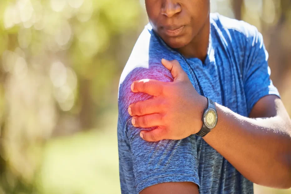
Glenohumeral ligaments serve as the dynamic stabilizers of the shoulder joint, and their restoration through open or arthroscopic procedures is vital to ensure shoulder function and prevent further instability.
The glenohumeral joint is supported by three nerves: suprascapular, lateral pectoral and axillary nerves as well as three synovial fluid-filled bursae: subdeltoid, subcoracoid and subcoracoid bursae.
Superior Glenohumeral Ligament (SGHL)
The glenohumeral labrum is an anatomically long crescentic structure extending from the anterior edge of the glenoid cavity to the posterior aspect of the humeral neck, serving to absorb, disperse and redirect tensile forces applied to the glenoid face. Additionally, its passive stabilization mechanism of shoulder is reinforced by its ligaments acting as static restraints against dislocation.
Three ligaments have been identified in the anterosuperior segment of the glenohumeral capsule: the superior glenohumeral labral ligament (SGHL), middle glenohumeral ligaments (MGHL), and inferior glenohumeral ligaments (IGHL). The SGHL begins its course from the upper pole of the glenoid labrum before passing into a groove between upper portion of lesser tubercle and biceps tendon, ventral to its origin.
As illustrated by MR arthrography (7a), the superior glenohumeral ligament forms a circumferential framework around the superior and anterosuperior portions of the glenoid labrum, creating a concave surface which articulates with the cartilaginous rim of the glenoid cavity. This connection is further secured by biceps tendon and rotator cuff, acting as dynamic restraints against humeral head displacement.
However, small areas of laxity in the SGHL and adjacent regions of the capsule are acceptable as this area does not undergo axial loading during abduction of the shoulder. As a result, an indirect vector of humeral head rotation transmits to the labrum, producing an SLAP lesion (see animation).
Most SLAP lesions involve the superior portion of the glenoid labrum, which is visible on MR images as an area of hypointensity that runs from glenohumeral ligaments to the biceps tendon. Some SLAP lesions present as an abnormality that spans from superior, anterosuperior, and anterior portions of the labrum; this may or may not be true in all instances. Radiologists must recognize the various forms of SLAP lesions to provide effective care; some affect only the anterosuperior labrum while others can extend into the MGHL and biceps tendon. Radiologists must recognize all variants of SLAP lesions so as to provide appropriate treatments, which include biceps tenodesis or tenotomy in case of SLAP III/IV lesions as well as selective resection of bucket handle (SGHL) lesions (SGHL lesions).
Inferior Glenohumeral Ligament (IGHL)
The IGHL is a hammock-like ligament connecting the humeral neck to the inferior glenohumeral labrum that serves as a critical passive stabilizing structure in shoulder stability. It often sustains damage from dislocations of anterior shoulder dislocations; additionally it may become injured from repetitive overhead activities or falls that exert rotational forces upon it.
This ligament can be divided into oblique and direct fibers. Oblique fibers attach to various locations along the humeral head, the superior glenohumeral cartilage, and proximal lesser tubercle of humerus while direct fibers run over an intra-articular portion of tendon of long head of biceps brachii to reach humeral semicircular ligament [3]. Furthermore, inferior glenohumeral labrum attachment to superior aspect of humeral capitellum by both types of fibers [IGHL].
Anterior inferior glenohumeral ligament (IGHL) injuries are one of the primary sources of shoulder instability, often as a result of trauma to an anterior shoulder dislocation and/or repeated anterior dislocations. They usually manifest themselves as Perthes lesions characterized by an anterior inferior labral tear with intact but stripped scapular periosteum and medial displacement of the IGHL relative to its position relative to the humeral head (Figure 6).
Biomechanical studies conducted on the IGHL have shown that both its superior band and anterior axillary pouch are capable of withstanding loads greater than 1000 N; the superior band’s insertion site may fail at lower strain rates while that of both its anterior axillary pouch and superior lobe remain resistant to failure at any strain rate.
MRI of the shoulder with fat-suppressed sequences can provide valuable insight into its ligamentous complex. When comparing findings of IGHL MRI findings with arthroscopic correlation, significant proportion of cases showed inferior capsular plicae, luxatio erecta humeri and superior labral avulsions on arthroscopic examination; while an IGHL mid-portion capsular tear was more likely present among patients who experienced multiple shoulder dislocations or small glenoid bone defects or disruption on MRA images.
Middle Glenohumeral Ligament (MGHL)
The middle glenohumeral ligament (MGHL) serves as a key stabilizer of the shoulder joint, preventing any anterior-inferior translation of the humerus against the glenoid cavity and arm adduction away from it. Many pathologies can impact it including shoulder dislocation, laxity of arm adduction laxity rotator cuff tears subscapularis tendon tears. Furthermore, its role is essential to normal and pathologic range of motion particularly during external rotation.
The MGHL connects to the anterior glenoid margin medial to the humeral head and spans across the rotator interval posterior to biceps tendon before attaching below SGHL attachment side; sometimes thickening into Buford complex.
MR imaging studies are often employed to examine the MGHL, with its appearance correlating with coexisting shoulder pathologies – in particular rotator cuff tears – but arthroscopic studies have also demonstrated that its structure may vary considerably: flat or leaf-like structures may exist alongside corded ones or even be entirely absent altogether.
Recent arthroscopic research conducted on healthy shoulders using the deltopectoral approach has demonstrated that the Medial Glenohumeral Ligament (MGHL) is an integral component of stabilization for the glenohumeral joint. It is reinforced by subscapularis tendon in the rotator interval and anchoring to both the humeral head at glenoid fossa and neck; and extends inferiorly and attaches to humeral condyles where it overlaps with superior and inferior glenohumeral ligaments.
The authors conducted a randomized controlled trial in which one group underwent standard arthroscopic procedure with an anterior portal while the other underwent an altered version that included both MGHL and subscapularis tendon / coracoacromial ligament (CHL) release techniques. Results revealed that both versions of the procedure produced similar clinical outcomes, suggesting that the MGHL is essential to maintaining stable adduction motion and should be repaired arthroscopically in patients with an Instability Severity Score less than 6, who qualify for surgery. Contrarily, patients who score 6 or higher require open surgery. The authors recommend that clinicians employ a standard instability score to determine if patients are suitable candidates for arthroscopic ligamentous repair; otherwise, consider their stability needs before making their decision about getting an MRI exam to better evaluate their shoulder.
Adduction Glenohumeral Ligament (AGHL)
The shoulder joint, otherwise known as the glenohumeral joint, is one of the most mobile joints in human anatomy. Unfortunately, this great mobility comes at the cost of decreased stability; most importantly, its primary pillar of support comes from ligaments and muscles of the rotator cuff, which provides multiple plane movement such as flexion/extension/abduction/adduction as well as external/internal rotation and circumduction.
The rotator cuff consists of four muscle tendons (superior supraspinatus, infraspinatus, teres minor, and subscapularis) which encase the humeral head and connect it to the scapula via the glenohumeral joint capsule. These tendon bundles encase the head in an almost continuous covering while their external surfaces blend in seamlessly; additionally the superior glenohumeral ligament (SGHL) and coracohumeral ligament (CHL) provide support on top surface of capsule.
Dense fibrous structure that connects the base of the coracoid process to both greater and lesser tuberosities of the humerus, as well as crossing from its anatomic neck through glenoid cavity to anatomic neck of humerus, merging with superior capsule and supraspinatus tendon superiorly. Proximal portion also envelopes Long Head Biceps Tendon’s insertion site within bicipital groove.
Rotator Cuff The rotator cuff extends laterally from the humerus and inserts at its anatomic neck just beneath the lesser tuberosity, with its anterior part attaching to the greater tubercle and limiting adduction while its posterior portion limits internal rotation.
Rotator Cuff Tendons Are Innervated By Suprascapular, Lateral Pectoral and Axillary Nerves The rotator cuff tendons are innervated by suprascapular, lateral pectoral and axillary nerves and are subjected to heavy strain during overhead activities, making them susceptible to injury and overuse. Overusing of the rotator cuff tendons may lead to impingement/subacromial bursitis where compressed tendons rub against the acromion of the scapula; patients typically present to primary care offices complaining of pain with overhead activity before physical therapy program usually resolves symptoms completely; should symptoms continue, evaluation by specialists is required.


